| Name | Haikouichthys ercaicunensis (“the fish of Haikou, from Ercaicun village”) |
| Classification | Phylum Chordata, family Myllokunmingiidae. A jawless fish (agnathan), regarded as among the oldest vertebrates |
| Age | Cambrian (about 518 million years ago) |
| Found in | Ercaicun village, Haikou, Xishan District, Kunming, Yunnan Province, China (the Chengjiang biota) |
| Length | About 2.5 cm |
| Described | 1999 (Luo, Hu & Shu) |
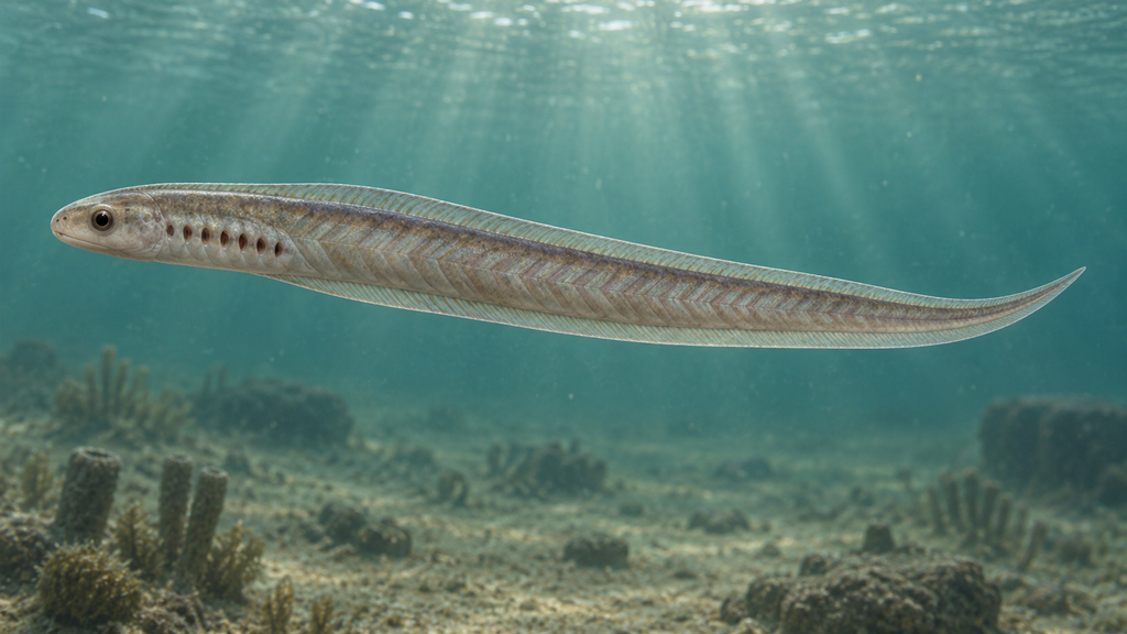
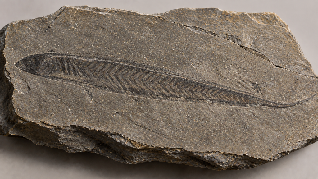
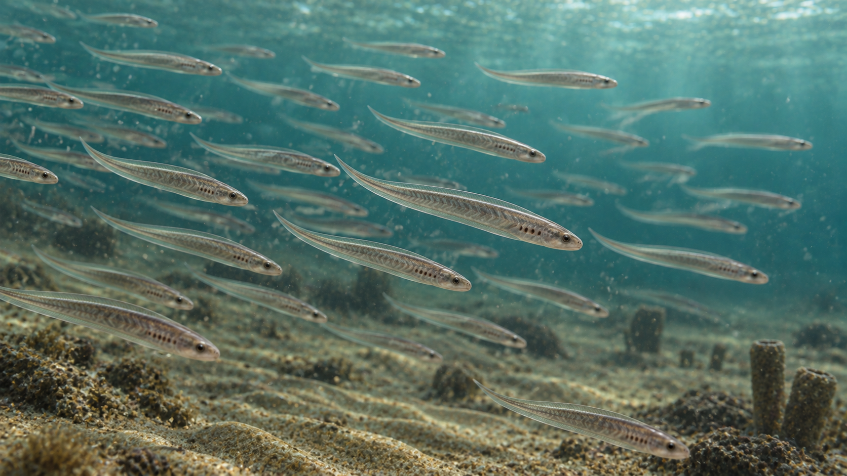
Haikouichthys was a small jawless fish that swam in the Cambrian sea. It had neither jaws nor paired fins; small vertebral elements were arranged along the notochord running down the center of the body, and V-shaped muscle blocks (myomeres) are marked along its sides. Among the oldest vertebrate fossils, it is often introduced in popular field guides and museums as the first fish.
Form and way of life
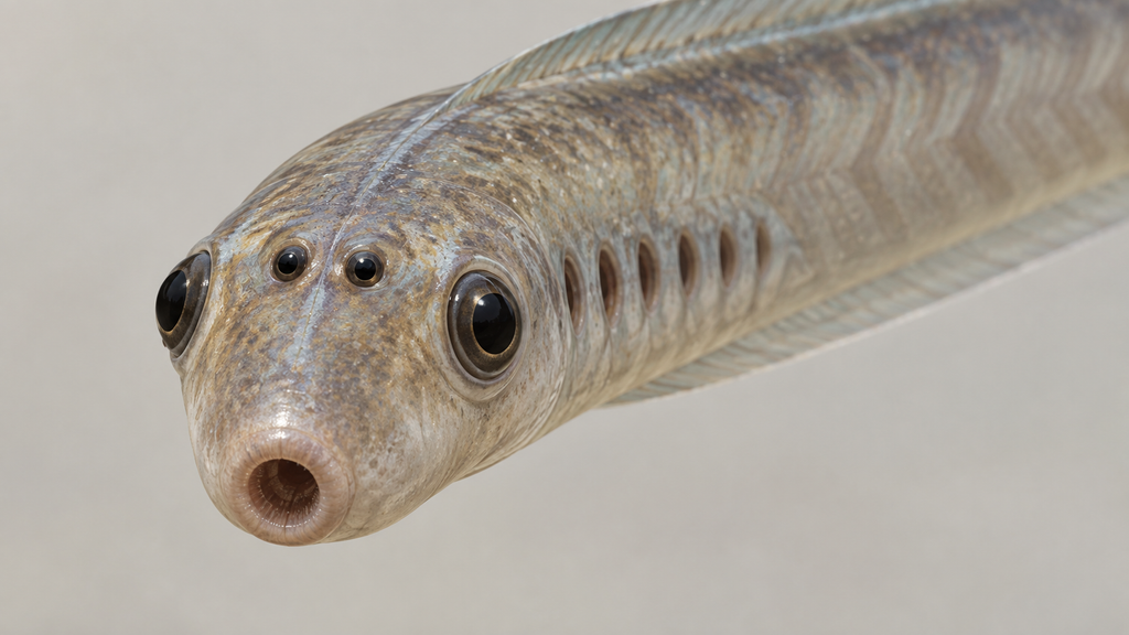
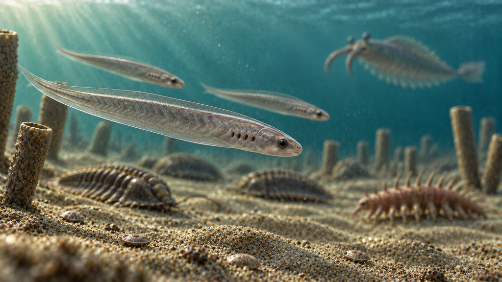
The body is slender, only about 2.5 cm long even as an adult. The region corresponding to the head bears a moderately large eye on either side, with a series of gills behind them. A single thin fin fold runs from the back along the tail and part of the underside. There are no pectoral or pelvic fins projecting to the sides, and the animal is thought to have swum by flexing its body.
With no jaws, the mouth was a structure that stayed circular and open — close in form to living lampreys and hagfish, and unable to bite large prey. It presumably took in organic matter drifting in the water and small plankton.
The notochord is preserved inside the body, with small cartilaginous elements arranged along it. These are the prototype of a backbone. The muscles are arranged as V-shaped blocks front to back, and the shape of these myomeres has been used as one of the grounds for distinguishing it from the closely related Myllokunmingia.
The same strata yield the large predator Anomalocaris, trilobites, the spine-bearing Hallucigenia and a variety of sponges. At a few centimeters long, Haikouichthys was among the small animals on the eaten side in this sea. A specimen of another myllokunmingiid species reported in 2026 preserves many individuals densely clustered on a single slab, raising the possibility that they swam in schools.
From one holotype to over 500 specimens
In 1999, a Chinese and British research team reported two fish-like animals from the Chengjiang biota: Myllokunmingia and Haikouichthys, both described as new species in the same paper. At that point, however, the basis for Haikouichthys was a single incompletely preserved specimen. Conclusions about the structure of the head and the internal anatomy were noted even then as provisional.
The situation changed in 2003. More than 500 specimens were assembled from a locality near Haikou, and the same team produced a fresh description. In over 300 specimens, a leaf-shaped projection under a millimeter across was confirmed at the front of the head, carrying the eyes, structures that appear to be nasal sacs, and a region that may be an otic capsule. The notochord with its associated vertebral elements, and the skeleton supporting the gills, were also clearly demonstrated at this point. The following year, 2004, a separate specimen was reported preserving the dorsal fin, ventral fold and tail together.
The question of the name, meanwhile, remains open. In 2002, a specimen combining the features of Haikouichthys and Myllokunmingia was found, and it was argued that the two are the same animal, the differences amounting to no more than variation in post-mortem decay and flattening. Because both species were published on the same day in the same paper, if they are merged, which name survives is left to the judgment of later researchers. Those taking this position use the name Myllokunmingia, and a 2017 monograph on the Chengjiang biota treated both under that name. Others hold that the differences in gill arrangement and myomere shape are real and keep them in separate genera. The matter is unresolved.
Another pair of eyes, on the midline
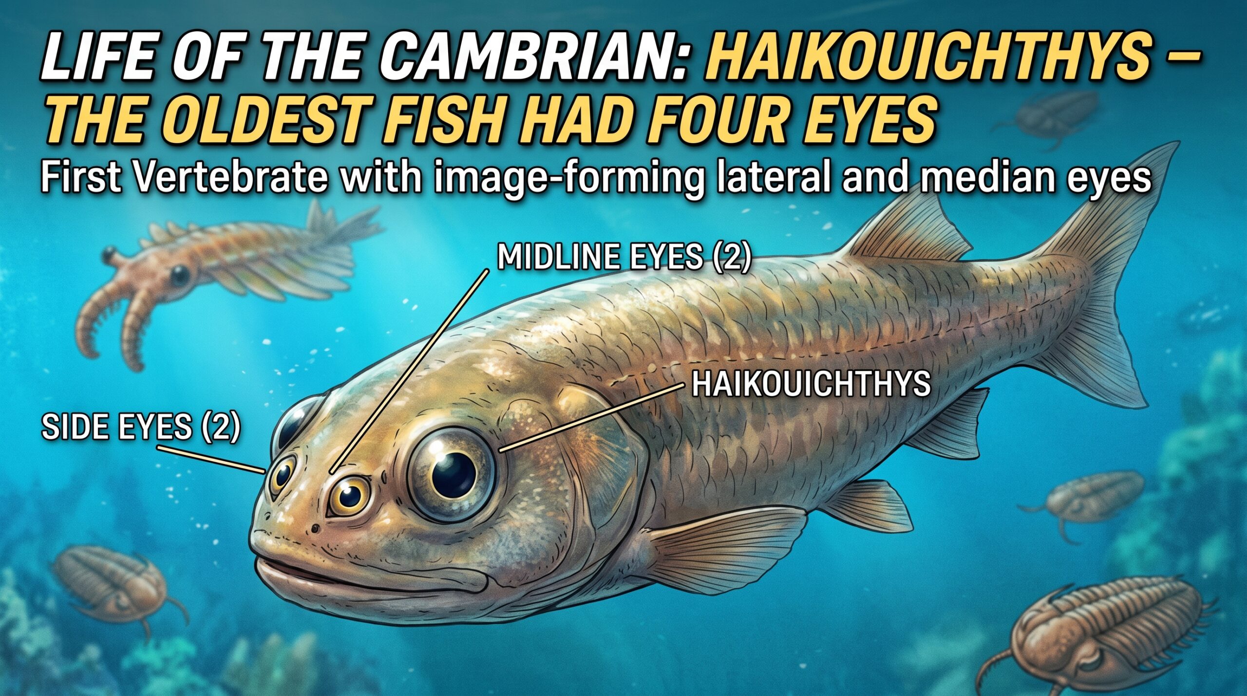
Since the 2003 description, it had been known that the head of Haikouichthys carries two small dark areas between the left and right eyes. These were long interpreted as nasal sacs for detecting smell.
In January 2026, a research team centered on Yunnan University published the results of re-examining those areas. The material was six specimens of Haikouichthys and four slabs of an unnamed myllokunmingiid species.
What they examined were melanosomes, the minute granules preserved in the fossils. These are intracellular sacs packed with melanin pigment, and within an eye they are concentrated in the pigment layer behind the retina. Granules extracted from the dark areas on the midline matched those from the lateral eyes of the same individual in both size and shape. Mass spectrometry and Raman spectroscopy further confirmed that the material is organic matter derived from melanin. Within the aggregation of granules there was also a regular ovoid structure, which the team judged to be a lens.
A pigment layer together with a lens means these two were also eyes capable of forming an image. Haikouichthys, in other words, had four eyes: one large pair on either side, and one small pair on the midline of the head.
From their position and form, the midline pair correspond to the pineal and parapineal organs of living vertebrates. In humans these have become small organs buried deep in the brain, releasing melatonin as darkness falls and regulating the rhythm of sleep. In lampreys and some lizards they can still sense light, and have long been called the third eye. This report sets out a line of descent in which that third eye was originally two, and in which they were true eyes able to form images rather than merely sense light.
The four eyes are thought to have covered different directions. A wide field of view including directly above would have helped both in detecting the approach of a predator such as Anomalocaris and in maintaining body orientation.
This is, however, an interpretation drawn from pigment structures preserved in flattened fossils. The work has been peer reviewed and published, but the reading of the structure identified as a lens, and whether the midline organs really formed images, are at the stage of awaiting testing against future specimens. Much remains unknown about this 2.5-centimeter animal with neither jaws nor bone.
Sources
The original description: Shu et al., “Lower Cambrian vertebrates from south China” (Nature 402, 42–46, 1999)
The redescription from over 500 specimens: Shu et al., “Head and backbone of the Early Cambrian vertebrate Haikouichthys” (Nature 421, 526–529, 2003) / PubMed
The argument that it is the same species as Myllokunmingia: Hou et al., “New evidence on the anatomy and phylogeny of the earliest vertebrates” (Proceedings of the Royal Society B 269, 1865–1869, 2002)
A specimen preserving the fins and tail: Zhang & Hou, “Evidence for a single median fin-fold and tail in the Lower Cambrian vertebrate, Haikouichthys ercaicunensis” (Journal of Evolutionary Biology 17, 1162–1166, 2004)
The report of four camera-type eyes: Lei et al., “Four camera-type eyes in the earliest vertebrates from the Cambrian Period” (Nature 650, 150–155, 2026) / the accompanying News & Views piece / coverage at Phys.org (free to read)
コメント