This article contains affiliate links. As an Amazon Associate I earn from qualifying purchases.

| Name | Lungfish, after the lungs they use to breathe air. The group’s scientific name, Dipnoi, is Greek for “two breaths”, referring to breathing with both gills and lungs |
| Classification | Vertebrates, bony fishes, lobe-finned fishes, lungfishes (six living species: one in Australia, one in South America and four in Africa) |
| Age | Devonian (about 400 million years ago) to the present |
| Where found | Fossils worldwide. The best-known fossil genus, Dipterus, comes from Middle Devonian lake deposits in northern Scotland. Living species inhabit rivers in south-eastern Queensland, Australia; the Amazon, Paraguay and lower Paraná basins of South America; and rivers, lakes and swamps of tropical Africa |
| Length | Largest is the African marbled lungfish (Protopterus aethiopicus), at about 2 m. The Australian lungfish (Neoceratodus forsteri) reaches about 1.5 m, the South American lungfish (Lepidosiren paradoxa) about 1.25 m |
| Weight | Over 40 kg in the Australian lungfish; up to about 17 kg in the marbled lungfish |
| Lifespan | At least 20–25 years in the Australian lungfish. One aquarium individual lived for more than 80 years |
| Genome size | About 91 billion base pairs in the South American lungfish, roughly 30 times the human genome and the largest animal genome sequenced so far |
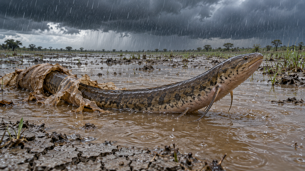
Lungfishes are lobe-finned fishes that have lungs as well as gills and gulp air at the surface. They appeared in the Devonian, spread through freshwater habitats around the world, and survive today as six species in Australia, South America and Africa. When their rivers and swamps dry out, the African species burrow into the mud, wrap themselves in a thin cocoon and sleep for months, sometimes years, until the water returns.
Appearance and Way of Life
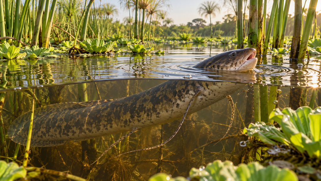
Living lungfishes come in two body plans. The Australian lungfish has a thick trunk covered in large, hard scales and fleshy, paddle-like pectoral and pelvic fins. Its dorsal, tail and anal fins merge into one, edging the rear half of the body. The five African and South American species are long and eel-like, with scales sunk into the skin and pectoral and pelvic fins reduced to thin filaments.
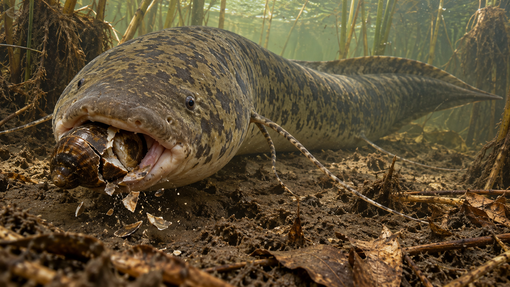
There are no rows of ordinary teeth in the mouth. Instead, the upper and lower jaws carry plates of fused teeth, called tooth plates, used to crush snails and crustaceans. The African species eat snails, frogs and fish, and also plant roots and seeds.
The lung shares its origin with the swim bladder of other fishes and opens into the back of the throat. The Australian lungfish has a single lung; the other five species have a pair. The Australian lungfish normally breathes with its gills alone and rises to the surface for air only when the river stagnates and the water runs low on oxygen. In the African and South American species, the gills shrink in adults, and most of their breathing depends on air. The South American lungfish drowns if it cannot reach the surface.
Lungfishes are the closest living fishes to the four-limbed vertebrates that moved onto land. The genome of the Australian lungfish, sequenced in 2021, confirmed that lungfishes are closer to tetrapods than the coelacanth is. That genome is about 14 times the size of ours, and around 90 percent of it is repetitive sequence. When the genomes of all six species were sequenced in 2024, the South American lungfish’s turned out to be more than twice as large again, at about 91 billion base pairs.
Fossil lungfishes were abundant in Devonian seas and lakes. Dipterus, from Middle Devonian lake deposits in Scotland, was a spindle-shaped fish with two dorsal fins, and it already had tooth plates. Tooth plates of Ceratodus and its relatives turn up in Mesozoic rocks in many parts of the world. The Australian lungfish, which lives in Queensland rivers, was described in 1870 under the name Ceratodus forsteri, placing it in that fossil genus.
A Living Cocoon in the Mud of a Dried-Up Swamp
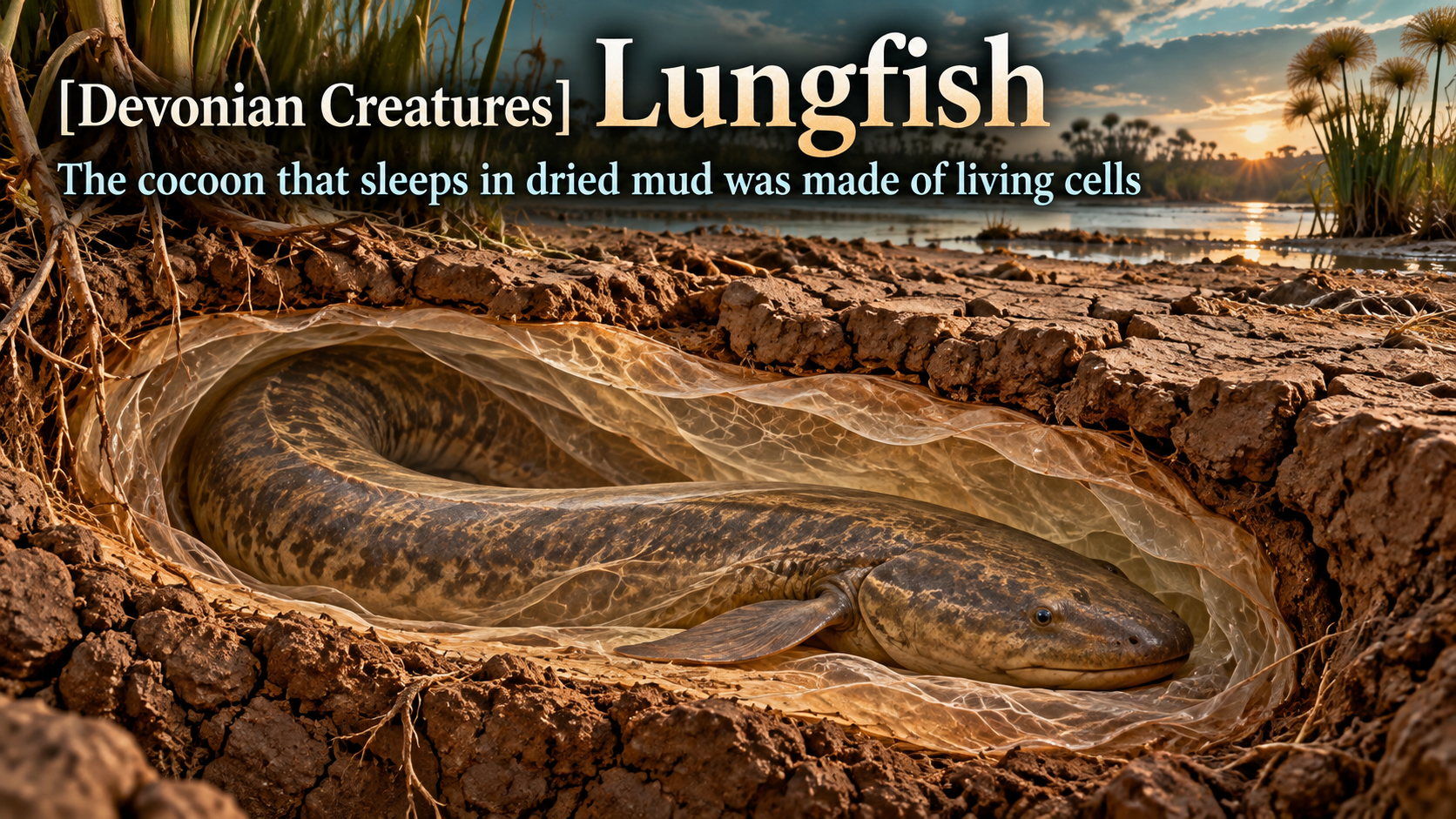
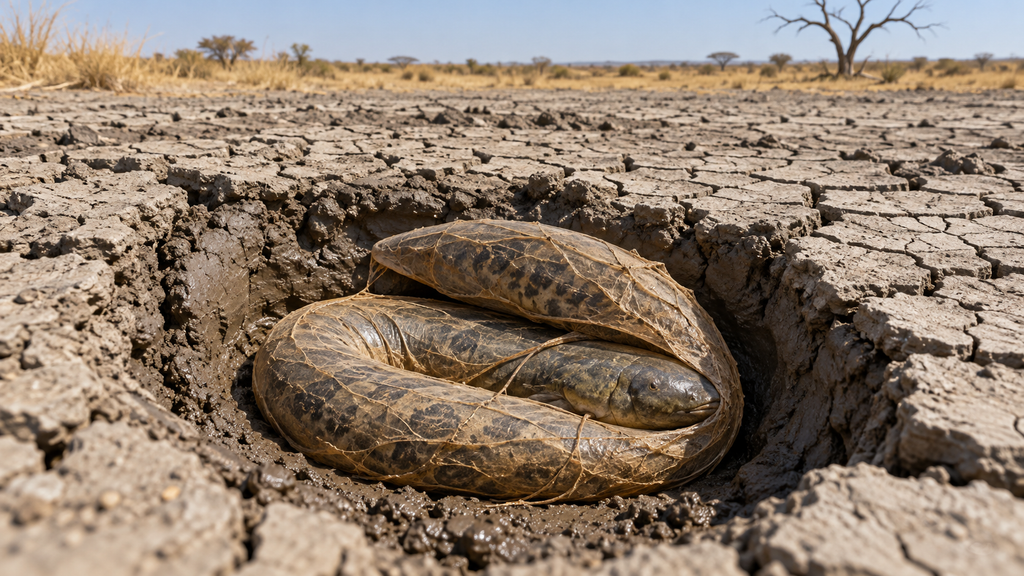
The swamps and river margins where African lungfishes live include places that lose all their water in the dry season. As the water recedes, the lungfish burrows into soft mud, bends its body and secretes large amounts of mucus from its skin. In the West African lungfish (Protopterus annectens), this mucus hardens within six to eight days into a thin brown cocoon around the body. Inside it, the fish eats nothing, barely moves, and keeps breathing air.
This state is called aestivation. In the wild it normally lasts seven to eight months, and during long droughts up to about four years; in captivity, a lungfish has been reported to emerge from its cocoon after seven years. Body functions slow sharply. Toxic ammonia produced in the body is converted into less toxic urea and stored, then flushed out when the rains return and the fish is back in water.
For a long time the cocoon itself was thought to be a dried, hardened layer of mucus. In 2021, Ryan Heimroth, Irene Salinas and colleagues at the University of New Mexico induced aestivation in an African lungfish (Protopterus dolloi) in the laboratory and examined the cocoons with microscopy and gene-expression analysis. The cocoons were 0.35 to 0.9 mm thick, made of many layers formed as the skin surface repeatedly shed and regenerated, and packed with living cells. Only about 7 percent of the cells were dead.
The cocoon also contained granulocytes, a type of white blood cell, that had migrated from the body. Granulocytes can release their own DNA as a net that entangles bacteria, known as an extracellular trap, and inside the cocoon bacteria were caught in these nets. When the team sprayed a DNA-digesting enzyme onto the skin of aestivating lungfish to destroy the nets, the cocoon kept its shape, but bacteria passed through it to the skin. The fish developed skin lesions and haemorrhages, and bacteria entered the bloodstream. The team concluded that the cocoon is a living tissue that protects the body from bacteria in the mud. The study, however, covered only the first two weeks of aestivation, and how well the cocoon protects the fish over an aestivation lasting years is unknown.
In 2024, Fernanda Palominos and colleagues at the University of California, Berkeley, posted the results of their own study of cocoons in the West African lungfish as a preprint, a paper not yet peer reviewed. It confirmed that the inner and outer layers of the cocoon consist mainly of living cells. The cocoon tissue, however, showed no activity against E. coli, and within about two to three weeks mould grew on the outside of every fish’s cocoon. The mould did not reach the fish inside. Both studies agree that the cocoon is living tissue; whether its job is to kill bacteria or to act as a physical barrier remains unsettled.
The habit of burrowing into mud to wait out the dry season is preserved in fossils too. Early Permian rocks in Texas and Oklahoma contain many vertical, cylindrical burrow casts, and inside them lie the bones of the lungfish Gnathorhiza, still in an aestivating position. The inner chamber of these burrows ranges from about 1.5 to 10 cm across and reaches up to about 50 cm in length. The living Australian lungfish, by contrast, cannot aestivate and does not survive once the water is gone. Among lungfishes, only the four African species pass the dry season sealed in a cocoon; the South American species shelters in a mud burrow and lowers its metabolism.
Related products
You can find books and products related to this article on Amazon.
If you buy through the links above, this blog may earn a commission.
Sources
Heimroth, R. D., Casadei, E., Benedicenti, O. et al. (2021). The lungfish cocoon is a living tissue with antimicrobial functions. Science Advances 7, eabj0829. https://doi.org/10.1126/sciadv.abj0829
Palominos, M. F. et al. (2024). The West African lungfish secretes a living cocoon during aestivation with uncertain antimicrobial function. bioRxiv (preprint). https://doi.org/10.1101/2024.07.05.602297
Chng, Y. R., Ong, J. L. Y., Ching, B. et al. (2016). Molecular Characterization of Aquaporin 1 and Aquaporin 3 from the Gills of the African Lungfish, Protopterus annectens, and Changes in Their Branchial mRNA Expression Levels and Protein Abundance during Three Phases of Aestivation. Frontiers in Physiology 7, 532. https://doi.org/10.3389/fphys.2016.00532
Hiong, K. C., Ip, Y. K., Wong, W. P. & Chew, S. F. (2013). Differential Gene Expression in the Brain of the African Lungfish, Protopterus annectens, after Six Days or Six Months of Aestivation in Air. PLoS ONE 8, e71205. https://doi.org/10.1371/journal.pone.0071205
Meyer, A., Schloissnig, S., Franchini, P. et al. (2021). Giant lungfish genome elucidates the conquest of land by vertebrates. Nature 590, 284–289. https://doi.org/10.1038/s41586-021-03198-8
Schartl, M., Woltering, J. M., Irisarri, I. et al. (2024). The genomes of all lungfish inform on genome expansion and tetrapod evolution. Nature 634, 96–103. https://doi.org/10.1038/s41586-024-07830-1
Olson, E. C. & Bolles, K. (1975). Permo-Carboniferous Fresh Water Burrows. Fieldiana: Geology 33(15). https://archive.org/details/permocarbonifero3315olso
Australian Museum. Australian Lungfish, Neoceratodus forsteri (Krefft, 1870). https://australian.museum/learn/animals/fishes/australian-lungfish-neoceratodus-forsteri-krefft-1870/
FishBase. Lepidosiren paradoxa. https://www.fishbase.se/summary/Lepidosiren-paradoxa.html
FishBase. Protopterus aethiopicus. https://www.fishbase.se/summary/Protopterus-aethiopicus.html
FishBase. Neoceratodus forsteri. https://www.fishbase.se/summary/Neoceratodus-forsteri.html

コメント